Reem Saadi Khalid1,
ABM Helaluddin1 ![]() ,
Mohamed Alaama1,
Abdualrahman M Abdualkader1,
Abdulrazak Kasmuri2,
Syed Atif Abbas3
,
Mohamed Alaama1,
Abdualrahman M Abdualkader1,
Abdulrazak Kasmuri2,
Syed Atif Abbas3
For correspondence:- ABM Helaluddin Email: abmhelal@iium.edu.my
Received: 21 April 2016 Accepted: 19 August 2016 Published: 30 September 2016
Citation: Khalid RS, Helaluddin A, Alaama M, Abdualkader AM, Kasmuri A, Abbas SA. Reliability of graphite furnace atomic absorption spectrometry as alternative method for trace analysis of arsenic in natural medicinal products. Trop J Pharm Res 2016; 15(9):1967-1972 doi: 10.4314/tjpr.v15i9.22
© 2016 The authors.
This is an Open Access article that uses a funding model which does not charge readers or their institutions for access and distributed under the terms of the Creative Commons Attribution License (http://creativecommons.org/licenses/by/4.0) and the Budapest Open Access Initiative (http://www.budapestopenaccessinitiative.org/read), which permit unrestricted use, distribution, and reproduction in any medium, provided the original work is properly credited..
Purpose: To evaluate the comparative efficiency of graphite furnace atomic absorption spectrometry (GFAAS) and hydride generation atomic absorption spectrometry (HGAAS) for trace analysis of arsenic (As) in natural herbal products (NHPs).
Method: Arsenic analysis in natural herbal products and standard reference material was conducted using atomic absorption spectrometry (AAS), namely, hydride generation AAS (HGAAS) and graphite furnace (GFAAS). The samples were digested with HNO3–H2O2 in a ratio of 4:1 using microwave-assisted acid digestion. The methods were validated with the aid of the standard reference material 1515 Apple Leaves (SRM) from NIST
Results: Mean recovery of three different samples of NHPs, using HGAAS and GFAAS, ranged from 89.3 - 91.4 %, and 91.7 - 93.0 %, respectively. The difference between the two methods was insignificant. A (P= 0.5), B (P=0.4) and C (P=0.88) Relative standard deviation (RSD)
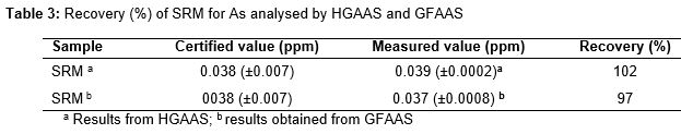
RSD, i.e., precision was 2.5 - 6.5 % and 2.3 - 6.7 % using HGAAS and GFAAS techniques, respectively. Recovery of arsenic in SRM was 98 and 102 % by GFAAS and HGAAS, respectively.
Conclusion: GFAAS demonstrates acceptable levels of precision and accuracy. Both techniques possess comparable accuracy and repeatability. Thus, the two methods are recommended as an alternative approach for trace analysis of arsenic in natural herbal products.
Introduction
Arsenic (As) is a hazardous element that occurs in trace amounts in various environmental samples due to both natural causes and anthropogenic activities [1]. Arsenic shows both metal and non-metal characteristics and has therefore been classified as a metalloid. Within the environment, arsenic exists in organic and inorganic forms in different oxidation states. Among them, arsenite trivalent [As3+] has a higher potential of toxicity than the arsenate pentavalent [As5+] [2].
Natural herbal products (NHPs) used for medical purposes are of great importance worldwide [3].The safety of NHPs is a vital issue for insuring public health. Arsenic contamination in NHPs has been reported globally [4-6]. Such contamination either originates from the raw materials themselves or released during the manufacturing process [7]. Consumption of contaminated products is encountering serious health risks. Symptoms of acute exposure to As include nausea, gastrointestinal distress and diarrhoea. Long term exposure to arsenic is a matter of concern mostly due its carcinogenic effects. Inorganic arsenic is known as a human carcinogen. Arsenic compounds can promote tumors in various organs such as liver, prostate, kidney [8]. Regular monitoring of such contaminant in NHPs requires simple and accurate analytical method.
Atomic absorption spectroscopy has been widely used for elemental determination at trace concentration levels [9]. Hydride generation-atomic absorption spectroscopy (HG-AAS) is routinely used for the determination of As at trace concentration levels in various matrices [10]. However, this method consumes relatively large quantity of chemicals and requires several steps for standards and samples preparation each step has significant impact on the accuracy and precision of the results [11]. Graphite furnace (GFAAS) has been efficiently applied for metals measurements at low concentration levels after reducing the interference problems by various techniques [12]. Therefore, this study aims to evaluate the extent of reliability of GFAAS as an alternative detection method to HG-AAS for As analysis in natural herbal medicinal products by comparing the level accuracy and precision between HGAAS and GFAAS.
Methods
Instrumentation
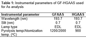
Perkin Elmer Atomic Absorption Spectrometer (AAS) A Analyst 800 equipped with EDL arsenic lamp. Autosampler graphite furnace (GFAAS) equipped with Zeeman effect background correction. Hydride generation (HGAAS) technique based on the reaction of acidified aqueous samples with a reducing agent to generate volatile hydrides that transport to the quartz cell atomizer by means of argon carrier gas. shows the instrumental parameters of GFAAS and HGAAS for As analysis.
Perkin Elmer Titan MPS 16 position vessels digestion system (microwave heating program) was used for samples digestion. Operating temperature from ambient to 170 °C and recommended pressure of 40 bars.
Chemical reagents and glassware
All chemicals and reagents used in this study were of analytical and trace metal grades. Trace metal grades 65 % HNO3 and 30 % hydrogen peroxide (H2O2) were obtained from Fisher Malaysia. Stock standard solution of inorganic arsenic (As) with a concentration of 1000 ppm, matrix modifiers palladium nitrate (Pd (NO3)2) and magnesium nitrate (Mg (NO3)2) were supplied by Perkin Elmer USA. Deionized water was used throughout the study. Sodium borohydride (NaBH4), sodium hydroxide (NaOH), L-ascorbic acid (C6H8O6), and potassium iodide (KI) were obtained from Merck (Germany). A standard reference material (SRM) 1515 apple leaves was obtained from the National Institute of Standards and Technology (NIST, USA). All glassware were soaked in 5 % (v/v) HNO3 overnight then rinsed with deionized water and dried using lab dryer FDD 720 prior to use.
Sample preparation
Natural herbal products originated from specific medicinal plants Eurycoma longifolia Tongkat Ali and Labisia pumila Kacip Fatimah in capsule dosage form were purchased from herbal medicine stores from various regions of Peninsular Malaysia.
An amount (0.5 g) of each sample was accurately weighed and placed in 75 ml vessel and then 5 ml of acid mixture of HNO3:H2O2 in a ratio of 4:1 was added. Samples were spiked with known concentration of As standard solution prior to digestion. The vessels were covered and heated to the target temperature of 170 oC for 10 - 15 min time was required for the digestion. The vessels were removed from the microwave rotor and allowed to cool to room temperature then cautiously opened in a fume hood and the inner walls were rinsed with DI water. The final volume of each sample was made up to 50 mL with DI water [13]. In HGAAS analytical technique samples were pre-reduced from arsenate pentavalent (V) to arsenite trivalent (III) state by adding 5 % w/v KI, 5 % w/v ascorbic acid and 10 % HCl, and the treated samples were allowed to stand at room temperature for approximately 40 min prior to analysis.
Spike recovery (R) was computed as In Eq 1.
R (%) = {(Co – Cr)/Ca}100 ……………………. (1)
Where Co, Cr and Cr are the concentration obtained, original concentration and analyte concentration, respectively.
Relative standard deviation was determined as the ratio standard deviation to the mean, expressed as a percentage, while recovery of SRM was calculated as the ratio of the measured concentration (HGAAS or GFAAS) to the certified value, expressed as a percentage
Statistical analysis
The data are expressed as mean ± standard deviation (SD, n = 6)) were evaluated by independent sample t-test using Statistical Package for the Social Sciences (SPSS). P < 0.05 was set as the level of significance.
Results
Methods comparison and validation
In this work basic analytical parameters were measured for GFAAS and HGAAS methods for arsenic analysis such as linear range, coefficients of correlation, LOD and LOQ. Calibration curves were constructed for each method in accordance with sensitivity check or the characteristic mass values recommended by the manufacturer. The LOD value was first calculated based on three times standard deviation (SD) for 10 replicates of the blank then the standard deviation of seven replicates of reagent blank spiked with known concentration of As standard solution was multiplied by the Student’s t-value to get the LOD value for As while LOQ value was calculated multiplying LOD by 10 [14]. The calibration ranges for HGAAS and GFAAS were 2 - 10 ppb. The coefficients of correlation for HGAAS and GFAAS techniques were 0.998 and 0.999 respectively. The LOD for HGAAS and GFAAS were 0.11 and 0.12 ppb while the LOQ were 1.1 and 1.2 ppb respectively.
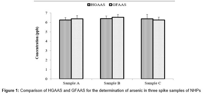
Accuracy and precision of HGAAS and GFAAS were compared by spike and recovery experiments using three different real samples of NHPs namely A, B and C to evaluate the sample matrix effect on the analytical systems. Samples were spiked with known concentration (7 ppb) of arsenic standard solution prior to microwave assisted acid digestion. The spike samples were analysed by HGAAS and GFAAS to measure the concentration of As analyte recovered as well as the level of the precision by measuring the SD and/or RSD.
The data (mean of six replicates ±SD) were analyzed using independent samples t-test to compare the recovery values of arsenic obtained from HGAAS and GFAAS. shows that there is no significant difference between two techniques.
The accuracy of analytical techniques was confirmed by the analysis of standard reference material 1515 apple leaves (SRM) from NIST. shows the result of SRM percentage recovery of As analysed by HGAAS and HGAAS.
Discussion
Generally, arsenic occurs at trace concentration levels in various environmental samples including NHPs [4,16]. The complex matrix of NHPs and low contraction level of As in such matrix require adequate analytical method to achieve good quality of analytical data. HGAAS is an accurate robust atomization technique for measuring hydride forming elements at low concentration level. It possesses the advantages of high sensitivity and high tolerance to interferences due to total separation of the analyte as a hydride from the matrix during the atomization stage. However, HGAAS is prone to drawbacks, as it requires tedious procedure for standards and samples preparations and involves hazardous materials. Various technical factors may also influence the hydride forming and the subsequent analytical results, such as tubing condition, valence state of the analyte, quartz cell position, and temperature [11]. GFAAS technique had a substantial improvement in analytical sensitivity after the introduction of matrix modifiers Pd-Mg salts. GFAAS has good detection limits for a majority of elements, with a small sample size for analysis 20 µl and minimum requirements for sample preparation [17].
The data for both methods demonstrated good linearity, similar linear ranges relevant to the low level of arsenic concentration in NHPs, and convergent values for LOD and LOQ values. This indicates good sensitivity at trace level of As analysis using HGAAS and HGAAS.
Results obtained from comparison experiments comparing both analytical techniques using spike matrices techniques for three different samples of NHPs as presented in table 2 show convergent recovery values of As in NHPs samples, namely A, B, and C, as measured by both techniques. Arsenic concentrations were compared by independent samples paired t-test using SPSS for each sample. No significant difference was observed between the methods. Nevertheless, GFAAS yielded higher recovery for As in samples A and B, which may be due to the effect of modifiers during the pyrolysis stage by reducing analyte loss and subsequently enhancing the sensitivity of the method. The precision of HGAAS and GFAAS was was within the acceptable values for repeatability of trace analysis [15]. Analysis of 1515 apple leaves (SRM) from NIST using HGAAS and GFAAS techniques confirmed the accuracy of both techniques.
The above results indicate that GFAAS and HGAAS methods possess comparable levels of accuracy and precision. The main advantage of GFAAS technique is the ability to conduct direct analysis with fewer sample pre-treatment requirements.
GFAAS technique has been developed for arsenic analysis in various samples of edible oil. Excellent recovery values were obtained for spike samples of different types of edible oil samples [18]. Emilene and co-authors developed HG-GFAAS detection method for arsenic determination in gasoline samples [19]. In a comparison study between GFAAS and HGAAS for arsenic analysis in different tissues (leg, breast, liver, and heart) of broiler chicken samples, the authors reported a lower detection limit for GFAAS, and adequate level of precision and accuracy in certified reference material and broiler chicken samples compared to HGAAS [20]. Results of the relevant studies have indicated that GFAAS offers sufficient levels of accuracy and precision, which is in agreement with the present study findings.
Conclusion
Both techniques demonstrate high levels of accuracy and precision for As trace analysis in NHPs. Natural products of therapeutic interest are being consumed increasingly worldwide. Thus, the developed GFAAS technique is a suitable, simple and less hazardous instrumental analytical method for regular monitoring of As to ensure the safety of NHPs and minimize toxicity.
Declarations
Acknowledgement
References
Archives


News Updates